FREQUENCY
The APERT syndrome also known as acrocephalosyndactyly type I (ACS1) is one of the most severe skull syndromes and occurs at a frequency 1-65.000 up to 1-88.000 births. It is characterized by premature fusion of certain skull bones (craniosynostoses). This early fusion prevents the skull from growing normally and affects the shape of the head and face. The craniosynostoses are responsible for the characteristic appearance of this syndrome.
CLINICAL PICTURE
The appearance of children with Apert syndrome is typical.
- The skull presents a reduced anteroposterior diameter (brachycephaly). The cranial vault often shows bone deficits.
- The head is long with a high forehead. The eye sockets are shallow and somewhat remote from one another (Πλατυπροσωπία - Ocular Hypertelorism).
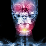
- The development of the face’s middle tertile is significantly weaker and this is a cause of serious respiratory obstruction (obstructive sleep apnea).
- The nose has a characteristic rounded appearance and it is flattened with low bridge.
- Teething presents serious disorders, with congestion and irregular arrangement of the teeth.
- The palate is vaulted and quite often there is a cleft.
The hands and feet present malfunction with abnormal development of multiple joints leading to their dysfunction as well as to severe syndactylies between the fingers and toes. Another problem in Apert syndrome is the continuous deterioration of the clinical picture that is observed with the progress of development.


ETIOLOGY
Two mutations in the FGFR2 gene of chromosome 10 are responsible for Apert syndrome. This gene produces a protein called fibroblast growth factor receptor 2. Among its multiple functions, this protein signals immature cells to become bone cells during embryonic development. A mutation in a specific part of the FGFR2 gene alters the protein and causes prolonged signaling, which can promote the premature fusion of bones and cartilages in the skull, hands, and feet. The mutations can be inherited from the parents or created without pre-existing family history. Most of the cases are caused by new mutations..
FUNCTIONAL PROBLEMS
The most serious functional problems in Apert syndrome are the following:
- Obstructive apnea, especially during sleep (nocturnal obstructive apnea)
- Increased intracranial pressure which may occur at any stage and age (see section). The increased intracranial pressure may be generalized (and therefore is easily detected) or be exerted only local pressure in the frontal lobes due to brachycephaly.
- Eye problems that often lead to reduced vision.
- Mental retardation that is observed in all children with Apert syndrome, but whose severity varies.
- Oily skin and acne.
- Frequent episodes of otitis.
- Less emphasis has been laid in the past to the disorders of the respiratory system. In addition to the throat narrowness there may be considerable problems in the larynx, the trachea and the bronchi, which can lead to serious respiratory problems.
- Finally, the characteristic of Apert syndrome is megalencephaly (or Macrencephaly abbreviated MEG) that is shown in some cases, i.e. the brain is abnormally large.
SURGICAL REHABILITATION
The surgical operations for the Apert syndrome repair usually start from infancy.
Depending on the findings, it may need:
- Incision in the posterior part of the skull (occipitoparietal area) in order to allow the brain additional room to grow when there are local pressure points.
- Incision and forward displacement of the anterior segment of the skull i.e. the orbitofrontal area in order to decompress the anterior segment of the brain and to improve exophthalmos.
At the age of ten, it usually needs a larger operation the so-called fronto-facial advancement i.e. the separation of the anterior part of the skull with the middle tertile of the face and their displacement forward. The surgery is intended to repair the shortness of skull and face, but also to relieve the obstructive apnea, which there is always to some extent. This surgery is possible to be performed in infancy as well, only on emergency cases if the obstructive apnea is severe and cannot be treated differently.
Simultaneously with the onset of the craniofacial complex repair in Apert syndrome it should begin the rehabilitation of the hands so the child's hands to gain full functionality. This is performed by specialized hand surgeons. Usually, the initial surgeries consist in release of the thumb and gradually the remaining fingers.