How does radiological imaging help to the diagnosis of craniofacial malformations?
The diagnosis of craniofacial malformations starts from the clinical examination that is performed by the craniofacial surgeon. At the end of the clinical examination it is likely to ask the patient to make a CT or MRI. This happens because these tests give the doctor the opportunity to assess in detail the skeleton of the skull and face as well as the central nervous system (CNS). More specifically:
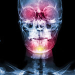
- A CT scan shows the skeletal molecules, aiding a detailed assessment of the craniofacial skeleton morphology.
- An MRI shows the soft tissues, presenting analytically the state of the brain.
In a second phase, if the surgical repair of the case is considered necessary, the imaging tests play an important role in the surgical planning.
What you should know before you make a CT or MRI scan:
- Both CT and MRI are not painful examinations. CT takes less than 1 minute, while the MRI takes about half an hour.
- In infants and children, general anesthesia is required in order not to move at all during the examination. (If there is some kind of movement during the scan, the image quality is altered)
- During CT, the patient is exposed to radiation, while in MRI not.
Note that:
- The radiation has been implicated for causing cancer and therefore every effort should be made to reduce the dose that gets into the child's organs. Make sure that the center where CT scans are applied follows strict protocols that prescribe low radiation doses for CT scans in children.
- With the use of low-dose radiation protocol, the radiation that the child takes is 20-fold reduced compared to an adult’s protocol.
- The radiation dose that the patient takes has been recorded by modern computed tomography machines.
- The factors that determine the radiation dose are the mA (milli Ampere) and the KV (kilo volt).
- The mA determines the number of photons while the KV their permeability.
- Reduction of mA and KV results in a drastic decrease of the radiation dose without diminishing the quality of bone imaging.
- Reduction of KV decreases the image quality of the soft tissues, something that is not so important since the soft tissues perfectly illustrated by MRI, which is usually done at the same date with the CT scan.
Do not forget:
- CT scan should not be applied unless it is absolutely necessary
- CT and MRI scan should always be done in consultation with the doctor that you have chosen for the surgical repair of the case. The doctor is responsible for deciding all the details.
- The clinical team and the staff of the radiological center must be in contact with the doctor about the details of each medical examination.
- You must always ask from the doctor the record of your examinations in CD, in order to allow their reproduction and their further processing with special software.