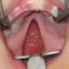
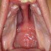
Palatoschisis or cleft palate is a split or opening in the roof of the mouth i.e. palate. The cleft palate may occur alone or in combination with cleft lip and alveolus. If the cleft palate is not accompanied by cleft lip, it is located in the middle line, whereas if it coexists with a cleft lip and alveolus, the cleft palate is located at the same side with cleft lip and alveolus.
A palate can involve two parts, the hard palate (the bony front portion of the roof of the mouth) and/or the soft palate (the soft back portion of the roof of the mouth). In the soft palate, there is a muscular layer that ensures its mobility which is essential for its proper functioning (speech, swallowing). It is understandable that the cleft palate ruptures the muscle layer and makes its normal functioning impossible.
The cleft palate may affect only the soft tissues or to be extended forward and include the hard palate bones. The breadth (width) of cleft palate varies widely, from too narrow, almost slit or very wide.
CLEFT PALATE REPAIR
The cleft palate repair is commonly performed at the age of 10-12 months and usually involves the repair of the soft and hard palate up to the alveolar area (where the teeth grow). But in the cases of wide cleft palates, the surgical repair (palatoplasty) is postponed for later time, without having any negative impact. The technique of cleft palate repair should be very delicate and cause the minimal injury to the soft tissues in order not to impair the dynamic growth of the upper jaw (maxilla).
During the cleft palate repair, it is necessary to repair the muscular layer accurately, in order to obtain the normal mobility of the soft palate.
The rehabilitation of the alveolar area is performed later, during the implementation of the bone graft. The technique of cleft palate repair plays a major role in the subsequent normal development of the upper jaw (maxilla). Poor technique can result in very serious disorders in the development of the upper jaw (maxilla).
EARS
Also, it is very important for a parent who has a child with cleft palate to know that there are great possibilities his/her child to have a particular sensitivity to the ears. More specifically, in children with cleft palate the Eustachian Tube (auditory tube or pharyngotympanic tube is a tube that links the nasopharynx to the middle ear) blocks having as a result fluid to be gathered in the middle ear. If this fluid is contaminated can cause acute otitis, which if not confronting effectively, could result in perforation in the tympanic membrane (or eardrum rupture). If the fluid remains in the ear for a long time it will become sticky and prevent the transfer of sound, resulting in damage to hearing. So, the continuous follow-up of the ears by a pediatrician or otolaryngologist is absolutely necessary.
In general, if it is found that the fluid remains and does not go away after treatment or if the child contracts otitis more than three times a year then it should be inserted ventilation tubes in the eardrums, through a very simple surgery, that will allow the free entry of air in the middle ear and prevent fluid collection. The repeated and continuous use of antibiotics not only must be taken as an alternative, but it is considered that it can have a harmful effect on the teeth, particularly in the permanent first molars, which may become loose and easily broken when the child bites something hard.
SPEECH
It’s obvious that the speech of a child with cleft palate that hasn’t been surgically repaired cannot be normal, as the speech development requires the integrity of the anatomic components of the mouth and pharynx (throat). The surgical repair of the cleft creates the necessary conditions for the proper speech development. In a small percentage, even after a cleft palate repair, speech problems may remain which initially have to be treated with speech therapy and if it’s necessary, with a small surgery that is called pharyngoplasty.
UPPER JAW (MAXILLA) DEVELOPMENT
In some cases, especially when there is cleft palate and alveolus, despite the compliance to the rules of the proper technique in cleft repair, the final development of the upper jaw (maxilla) lags behind the development of a normal jaw. This is due to that the dynamic of jaw growth can be reduced in cases of clefts. In this case, when the development is completed in the late teens, the upper jaw will be in the most rear position in relation to the lower jaw. This problem is repaired by an additional procedure (maxillary osteotomy), with which it is achieved the placing of the upper jaw in the correct position relative to the lower jaw (in severe cases, it is used the method of distraction osteogenesis) and thereby the cycle of rehabilitation surgeries is completed.


