The eye orbit is the part of the facial skeleton that contains the eye, the muscles that move it as well as the adipose tissue. The eye orbit consists of the skull and facial bones which effectively protect the eye from injuries.
EYE ORBIT AND EYE INJURIES
Despite the protection that the skeleton of the eye and the eyelids ensure to the eye, the eye is often subjected to injuries.
Orbital fractures may cause except from malformation and serious functional problems such as:
- Diplopia (or double vision) (i.e. the patient sees two images of one subject) due to the injury of the muscles that move the eye or due to eye displacement, or even vision loss.
INITIAL ASSESSMENT OF THE SITUATION
Immediately after the injury, the patient should be examined by
- Ophthalmologist (to assess the condition of the eye), and by
- Reconstructive surgeon (preferably with craniofacial surgery knowledge), to assess the damage of the orbit and eyelids.
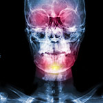
- For the better assessment of the damage, a 3D Reconstruction CT Scan may be needed to be performed.
REHABILITATION
In the rehabilitation of the orbital area traumas, priority has:
- The immediate rehabilitation of the eye
- The soft tissues and eyelids should be rehabilitated, as soon as the situation of the injured patient allows it, in order to protect the eye
- The rehabilitation of the skeleton which may be postponed as long as the oedema (swelling) subsides, unless the eye or the optic nerve of broken bone fragments is at risk